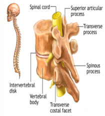
The spinal cord is a collection of nerves that travels from the bottom of the brain down your back. There are 31 pairs of nerves that leave the spinal cord and go to your arms, legs, chest and abdomen. These nerves allow your brain to give commands to your muscles and cause movements of your arms and legs.

The spinal cord is very sensitive to injury. Unlike other parts of your body, the spinal cord does not have the ability to repair itself if it is damaged. A spinal cord injury occurs when there is damage to the spinal cord either from trauma, loss of its normal blood supply, or compression from tumor or infection.
When spinal cord injuries occur in the neck area, symptoms can affect the arms, legs, and middle of the body. The symptoms may occur on one or both sides of the body. Symptoms can also include breathing difficulties from paralysis of the breathing muscles, if the injury is high up in the neck.
This type of cervical spine injury happens when the soft spinal discs bulge or rupture out of the spinal canal and put pressure on nearby nerve roots or the spinal cord. The culprit is usually some type of sudden force.
Over time, wear and tear on the cervical spine can injure it and cause the discs in the cervical spine to degenerate. The degeneration process can be exacerbated by a fall or twisting injury to the neck.
When spinal injuries occur at chest level, symptoms can affect the legs. Injuries to the cervical or high thoracic spinal cord may also result in blood pressure problems, abnormal sweating, and trouble maintaining normal body temperature.
The intervertebral joint is the joint that joins the levels of the spine together. Injury to this joint is usually due to a forced movement forward or backward of the thoracic spine. Pain can be felt locally about 2 cm to the side of the spine and may radiate around the chest wall to the front of the chest. Pain is increased with forward or backward movement of the spine.
This injury is common in many sports such as throwing sports, football, basketball and boxing. It is also commonly done when lifting heavy objects.
The costovertebral joint is a joint in the back that joins the rib and the spine. Common problems arise from spraining the joint during forced chest movement or from inflammation due to arthritis.
This injury is very common in contact sports such as football and rugby. It most commonly occurs as a result of a blow to the ribs.
This is the most common cause of pain in the thoracic spine in adolescents, especially boys. It is a hereditary back disease in which the back becomes rounded due to the bodies of the vertebrae becoming wedged shaped.
This is a curvature of the spine in a sideways direction which causes the spine to be S-shaped. Symptoms with scoliosis are not always present. Symptoms include complications due to muscle weakness and joint “looseness” on the convex side and muscle tightness and spasm with joint tightness on the concave side.
When spinal injuries occur at the lower back level, symptoms can affect one or both legs, as well as the muscles that control the bowels and bladder.
Low back pain can be caused by structures being too tight (hypo-mobility) or too loose (hypermobility). The pain-producing structures in the lumbar spine include the vertebra, the facet joints (links two vertebrae together in your spinal column), intervertebral disc, ligaments, nerves, and their protective coverings, muscles, and their attachments.
The intervertebral discs are composed of a soft, inner nucleus pulposus surrounded by a tough fibrous outer ring, the annulus fibrosus. With trauma and/or aging, the annulus fibrosus can weaken and thin (disc degeneration or herniation), particularly with the repetitive combination of bending forwards while rotating the trunk i.e. lifting.
LBP may also be caused by spondylolysis, or a stress fracture of the pars interarticularis, a region of the vertebra. This is often seen in sports involving repeated back extension and rotation, such as gymnastics, cricket fast bowling or tennis. While it was thought to be congenital, it is probably an acquired overuse injury. The fracture usually occurs on the opposite side to the one performing the task i.e. a left-sided fracture occurs in a right-handed tennis player.
Another commonly encountered cause of LBP is spinal canal stenosis. It is a condition that is rare in young and middle-aged athletes but may be seen occasionally in older athletes. The condition is caused by arthritic degeneration of the spine, resulting in the vertebra, facet joints, and ligaments that surround the spinal nerves of the spinal cord becoming enlarged. In this manner, these structures may compress one or several spinal nerves, causing LBP, leg pain, and leg numbness while walking.


Cancer Treatment in India, VSD Closure (Ventricular Septal Defect), ASD Closure Surgery (Atrial Septal Defect), Heart hole closure surgery in india, Brain Tumour Surgery in India, Craniotomy Surgery for Treatment of Brain Tumour in India, Brain Stem Glioma Treatment in India, Deep Brain Stimulation surgery in india , Best Spine Surgery In India , Cervical Spine Disorders Surgery and Treatment in india , epilepsy surgery in india , Heart Surgery in india , Best open heart surgery hospital in India , All about Spine Surgery, Types of Back Surgery , Top Doctors In India , Best Hospitals in India , Bhavin Desai cardiac surgeon, Best kidney transplant doctor in IndiaBest hospital for limb lengthening surgery in India, Best hospital for neurosurgery in India, Best hospital for bypass surgery in India, Narayana Hrudayalaya online appointment, Best orthopedic hospital in Bangalore, Best open heart surgery hospital in India, Best bypass surgeon in India, best spine surgery hospital in India, Artemis hospital Gurgaon, KD hospital medical appointment
Limb lengthening surgery cost in India, DSA test, Diabetes treatment in India